Which Best Describes the Blood Flowing in an Artery
It is oxygen rich B. Correct answer to the question Which best describes the blood flowing in an artery.

Pin On Body
B Arteries have a smooth muscle layer on the outside which provides some stiffness.

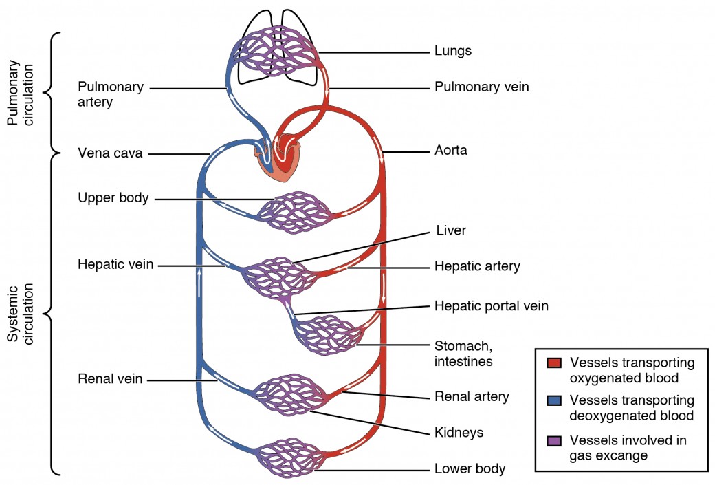
. Valves prevent the backflow of blood into the atria and ventricles. As the right ventricle contracts it sends oxygenated blood through the aorta to all tissues of the body. Large arteries are able to drive blood flow when the heart is relaxed because lymph Interstitial fluid proteins extravasated leukocytes dietary fats and even pathogens are components of the fluid in lymphatic vessels which is called.
It moves away from the heart. As arteries carry blood so it also plays an important role in delivering nutrients and removal of waste products like carbon dioxide. It moves toward the heart.
Blood is usually in laminar flow in the vessels. Capillaries provide blood to individual cells. Therefore lymph fluid flows freely from the tissue spaces into the bloodstream.
Which of the following routes of blood flow is correct. Lymph vessels have no valves. A All carry oxygenated blood to the heart.
The term viscosity describes the resistance to flow that arises because of the intermolecular attractions between fluid layers. Veins transfer oxygen-depleted blood away from the heart. C All contain valves to prevent the backflow of blood.
Deoxygenated blood flowing through the pulmonary veins is carried to the right atrium. Arteries transfer oxygen-rich blood to the heart. Because blood viscosity increases exponentially with increases in hematocrit the concentration of red blood.
Lymph flow is propelled by the contraction of the heart b. The quantitative arteriographic analyses were performed on. Heart- venule - medium vein- large vein - capillary - conducting artery - distributing artery - arteriole - heart B.
D Only large arteries are lined with endothelium. A Arteries have an endothelial lining that reduces friction and promotes smooth blood flow. D Valves along major arteries maintain directed blood flow.
B All carry blood away from the heart. A Lymph flow is propelled by the contraction of the heart B The flow of lymph is slow compared with that of the blood C One of the functions of the lymph is to absorb lipids from the biliary tract D Lymph vessels have no valves so there is a free flow of lymph fluid from the tissue spaces into the bloodstream. Arteries are also important in the circulation of immune cells in the body through blood.
Which statement best describes arteries. It is oxygen poor. One of the functions of the lymph is to absorb lipids from the biliary tract d.
Therefore the correct answer is c. It means you can feel the pulses of blood flowing through your main artery in the uterus uterine artery just by touching the area where the uterine artery passes through. A when pressure in the brachial artery is greater than in the blood pressure cuff and greater than the diastolic pressure B when pressure in the blood pressure cuff is less than the diastolic pressure.
When measuring blood pressure with a sphygmomanometer which of the following best describes when blood flow in the brachial artery is turbulent. Which statement best describes the function of veins arteries and capillaries in the circulatory system. Fluids with particularly strong intermolecular attractions offer a high resistance to flow and have high coefficients of viscosity.
The flow of lymph is slow compared with that of the blood c. Therefore it also helps in maintaining the pH of the blood. Thirty patients with an isolated lesion of the left anterior descending coronary artery LAD were studied by means of on-line quantitative coronary arteriography intracoronary Doppler flow velocity measurements and dipyridamole echocardiography 6 months after percutaneous transluminal coronary angioplasty.
When measuring blood pressure using a sphygmomanometer turbulent blood flow is seen to occur at the time when the blood cuff pressure is greater than the diastolic pressure in the brachial artery but. It contains no red blood cells. C Elastin fibers in arterial walls allow stretching of the arteries.
Heart - large vein - medium vein - venule - capillary - arteriole - distributing artery - conducting artery - heart.

Understanding Heart Disease Chart 22x28 Ischemic Heart Disease Heart Disease Heart Conditions

Pin On S C H O O L
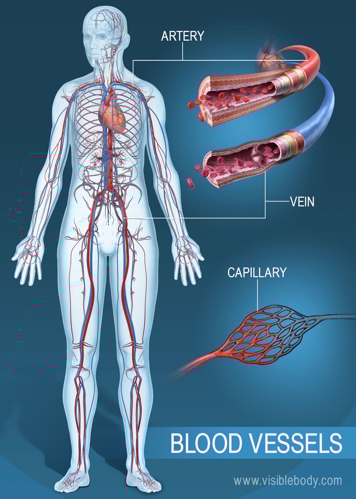
Blood Vessels Circulatory Anatomy

Yale School Of Medicine Neonatal Nurse Pediatric Nursing Midwifery Student
Structure And Function Of Blood Vessels Anatomy And Physiology Ii

Pin On Cardio

Pin By Samantha Rottman On Medical Heart Veins Medical Knowledge Good Health Tips Heart Surgeon

A Typical Blood Vessel A Shows A Normal Artery With Normal Blood Download Scientific Diagram
Blood Vessels Circulatory Anatomy

Anatomy Kidney Renal Artery Renal Vein And Ureter Anatomy Images Kidney Anatomy Kidney

Pin On Lymphatic System
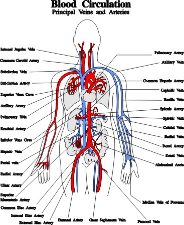
What Is The Correct Order Of Blood Flow Quora

Pin On Cvt

Pin On Salud

Pin On Interesting

Gross Anatomy Of The Kidney Anatomy And Physiology Openstax Renal Physiology Kidney Anatomy Renal
Blood Vessels Arteries Capillaries Veins Vena Cava Central Veins Lhsc

Pin On Health Is Wealth

Simple Diagram Of The Heart Heart Diagram Human Heart Diagram Simple Heart Diagram
